|
|
Epiglottitis
General Considerations
- Acute bacterial epiglottitis
- Life-threatening, medical emergency due to infection with edema of epiglottis and aryepiglottic folds
- Organism
- Introduction of Haemophilus influenzae type B vaccine in 1985 has led to marked decrease in number of cases of epiglottitis
- Still remains the most common cause
- Also caused by
- Pneumococcus
- Streptococcus group A
- Viral infection – herpes simplex 1 and parainfluenza
- May also be caused by thermal injury and angioneurotic edema
- Age
- Typically between 3-7 years
- Peak incidence has become older over last decade and is now closer to 6-7 years
- Location
- Purely supraglottic lesion
- Associated subglottic edema in 25%
- Associated swelling of aryepiglottic folds causes stridor
Clinical Findings
- Classical triad is: drooling, dysphagia and distress (respiratory)
- Abrupt onset of respiratory distress with inspiratory stridor
- Sore throat
- Severe dysphagia
- Older child may have neck extended and appear to be sniffing due to air hunger
- Resembles croup clinically, but think of epiglottitis if:
- Child can not breathe unless sitting up
- “Croup” appears to be worsening
- Child can not swallow saliva and drools (80%)
- Cough is unusual
Imaging Findings 
- Patient needs to be accompanied everywhere by a physician experienced in endotracheal intubation
- Imaging studies are not always necessary for the diagnosis and may be falsely negative in early stages
- Lateral radiograph should be taken in the erect position only, as
- Supine position may close off airway
- Enlargement of epiglottis
- Thickening of aryepiglottic folds
- Circumferential narrowing of subglottic portion of trachea during inspiration
- Ballooning of hypopharynx and pyriform sinuses
- Reversal of the normal lordotic curve of the cervical spine
- Fiberoptic-assisted, nasotracheal intubation is procedure of choice, so long as airway is secured
Differential Diagnosis
- Croup
- Dilatation of the hypopharynx
- Dilation of the laryngeal ventricle
- Narrowing of the subglottic trachea
- Epiglottis is normal
- Enlarged adenoids
- Compression of nasopharyngeal airway
- Frequently associate with enlargement of the lingual tonsils
- Epiglottis is normal
Treatment
- Secure airway
- May require intubation or emergency tracheostomy
- Some use IV steroids
- Empiric antibiotic therapy
Complications
- Danger of suffocation secondary to complete airway closure
- Pneumonia
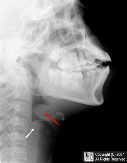
Epiglottitis. A lateral radiograph of the neck using soft tissue technique demonstrates an enlarged epiglottis (red arrow) with markedly thickened aryepiglottic fold (white arrow) diagnostic of acute epiglottitis.
For additional information about this disease, click on this icon if above.
For this same photo without the arrows, click here
|
|
|

)
)
)